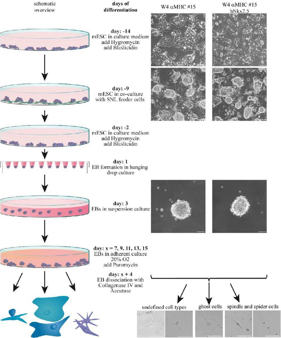
Fig. 3. Cardiogenic differentiation protocol of mESCs: Schematic overview of the cardiogenic differentiation protocol and representative cell pictures for murine ES cells using: W4, W4 αMHC and W4 αMHC hNkx2.5 clones. mESCs were cultivated in LIF supplemented medium with appropriate antibiotic pressure (W4 αMHC: hygromycin; W4 αMHC hNkx2.5: hygromycin and blasticidin). Afterwards, cells were co-cultured with mitotically inactivated murine STO cell line-derived SNL 76/7 (73). Prior to differentiation, cells were detached from feeder cells, and subsequently cultured for additional 2 days with appropriate antibiotic pressure. Specific cardiac differentiation was induced through EB formation and subsequent culturing in differentiation medium. αMHC-selection using puromycin was induced at various points in time (day: x = 7, 9, 11, 13, 15), respectively. A dissociation step 4 days after selection (day: x + 4) led to the generation of single cells with the following morphogenic classification: spindle and spider like cells, large round and large square cells or an undefined cell population including large longitudinal and small round cells. There are no living cells present in the W4 population after puromycin treatment scale bar: 100 µm.